Radiosurgery
Motivation

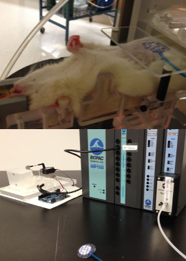
Fig. 1 (Upper) Rat in anesthesia cradle. (Lower) Pressure sensor and Arduino-driven breath amplifier-box.
Cardiac arrhythmia is a deadly condition in which one's heart beat becomes irregular and results in oxygen deprivation. The current treatment for atrial fibrilation (a type of cardiac arrhythmia) is cauterizing (burning) part of the heart so that scar tissue will electrically isolate the tissue generating the spurious electrical impulse.
Knowing that proton radiation kills cells and creates scar tissue, our research team was designing a non-invasive proton radiation treatment. I joined this project during the dose-finding portion of the study. We used a rat model to determine the radiation dose required to scar cardiac tissue. The treatment was to target the sinoatrial node, the heart's "spark plug," with radiation. To detect the development of scar tissue in the sinoatrial node we relied on an electro-cardiogram (ECG).
The animal procedure was :
sedating the rat,
giving the rat a CT-scan (3D x-ray),
planning a two-beam proton radiation treatment,
administering proton radiation, and
monitoring the heart for signs of scarring in the weeks that followed.
For the first step, Ying Nie, our fearless biologist, placed the conscious rat in an anesthesia box before comfortably securing the sedated rat in the half-pipe anesthesia cradle that would carry it for the duration of the experiment (see Fig. 1 upper).
The second step was administering a chest CT-scan to the rodent; the resulting 3D image gives our radiation specialist the information to create a two-beam radiation treatment plan. For a successful chest scan, the x-ray radiation must be gated (paused) during breathing to keep the exposure from blurring during chest movement. On human subjects, a box with reflective dots is placed on their chest and the CT-scanner registers its movement with a video feed. Unfortunately, this setup didn't work, because rats are too small, their breathing amplitude is too shallow, and were pointing away from the camera.
Our solution was to use a pressure sensor to measure the rat's breathing and artifically move the reflective box to interface with the CT scanner's existing video recognition system. The BIOPAC pressure sensor (Fig. 1 lower) output a 10 Volt DC analog signal, and I programmed an Arduino Uno to convert this to a pulse width modulation signal driving the servo motor which moved the reflective box. The first video gives an overview, and the second video shows it in action.
While the treatment plan (step three) was being created, I measured the spatial offset in the gantry 1 using proton-sensitive film. Then we performed step four: fixing the rat on the table, dialing in the offset, and administering the prescribed radiation dose.
The final step took place over the following weeks as a cardiologist monitored the rat's ECG for aberrations caused by scar tissue forming in the heart.
- 1
-
Proton gantry is a large machine designed to move a proton beam around a patient receiving radiation therapy.